

|
|
Structural and Conformational Analysis of Oligosaccharides by NMR |
 |
|||||||||||||
 |
NMR spectroscopy plays a critical role in the structural
and conformational analysis of oligosaccharides. In particular, chemical
shifts and spin-spin coupling constants are useful in determining the
composition of sugar residues and the positions and configurations of
glycosidic linkage. Vliegenthart and co-workers have so far expanded a
systematic study on 1H chemical shifts of oligosaccharides
and described numerous structures by using a “structural reporter
group”. Sugabase, a carbohydrate NMR database dealing with 1H
and 13C chemical shift values, is now available on the World
Wide Web. Consequently, it has become easier to identify oligosaccharides
with a common structural framework. Since NMR spectroscopy is inherently
insensitive, structural analysis with a small amount of sample is still
laborious. Recently, NMR sensitivity has been greatly improved by the
emergence of higher magnetic fields and cryogenic probes. Thus NMR spectroscopy
will no doubt continue to play an important role in the structural analysis
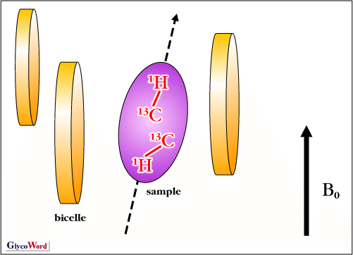
of oligosaccharides. NMR spectroscopy is of great use in the conformational analysis of oligosaccharides as well as the determination of covalent structures. Since oligosaccharides are not crystallizable in general, information provided by NMR spectroscopy on the conformation of oligosaccharides in solution at atomic resolution is quite important. Because individual sugar rings are usually fixed in the “chair” form, conformation of oligosaccharides is primarily characterized by the glycosidic torsion angles. In order to determine the glycosidic torsion angles, NOEs and spin-spin coupling constants, which are related to 1H-1H distances and dihedral angles, respectively, have so far been mainly used. Collection of enough number of restraints is so difficult that conformation of glycosidic linkage has often been described based on NMR data combined with theoretical calculations such as molecular dynamics simulations. In accordance with the recent development of stable isotope labeling techniques, novel NMR parameters that offer structural information have emerged. 13C-labeling of oligosaccharides has opened a new avenue to conformational analysis using 3JCH and 3JCC coupling constants, in addition to 3JHH, which define the torsion angles. Furthermore, at higher magnetic field, it becomes possible to observe residual dipolar couplings of oligosaccharide samples weakly oriented in the presence of ordering media (Figure 1). The magnitude of residual dipolar couplings depends on the distance between the two nuclei (e.g. pair of 13C and 1H) and the angle between the bond vector and the principal axis of the alignment tensor. Residual dipolar coupling is shown to be a useful NMR parameter for determining the conformation of oligosaccharides (1). NMR spectroscopy plays an important role in the conformational analysis of oligosaccharides bound to protein as well as free oligosaccharides. For instance, transferred NOE (trNOE) is used to determine the conformation of the bound oligosaccharides even though the conformational information of protein is absent. It is also possible to identify epitopes of oligosaccharides recognized by target proteins and to find an oligosaccharide ligand that is capable of interacting with a particular protein from the oligosaccharide mixture. Recently, it has been possible to analyze the glycoprotein itself, which was difficult previously (2)(3). By stable isotope labeling of glycans, it becomes feasible to elucidate the conformation and dynamics of glycans attached to proteins based on NMR data such as NOEs or relaxation rates. Conformational analyses of glycans attached to glycoprotein are very important for obtaining unique knowledge that has never been possible with liberated oligosaccharides and provide information regarding the structural basis and functions of glycans. |
|||||||||||||
 |
||||||||||||||
| Fig. 1 Schematic representation of the partial alignment of an anisotropic molecule labeled with 13C in the presence of disk-like micelles (bicelle). The bicelles align spontaneously with respect to the applied magnetic field (B0), and the molecule gains a small net degree of alignment and residual dipolar couplings can be measured. |
||||||||||||||
| Yoshiki Yamaguchi (Graduate School of Pharmaceutical Sciences, Nagoya City University) |
||||||||||||||
|
||||||||||||||
| Oct. 31, 2002 | ||||||||||||||
|
|
||||||||||||||
|
||||||||||||||