

|
|
Psychosine and Multinuclear Cell Formation in Globoid Cell Leukodystrophy |
 |
||||||||||
 |
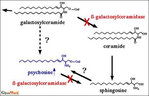
Globoid cell leukodystrophy (GLD) is a recessive autosomal disorder which is characterized by the reduction of beta-galactosylceramidase (GALC) activity. GALC hydrolyzes beta-galactosylceramide (GalCer) to galactose and ceramide. The symptoms of GLD are exclusively neurological (myotonia, seizure, etc.). Since GalCer is abundant in oligodendrocytes which maintain the myelin sheath functions, the abnormal metabolism of GalCer causes loss of function of myelin. The neurological symptoms in GLD are attributed to the loss of function of myelin. GalCer, however, does not accumulate in the patient's brain despite the sluggish GALC activity, but psychosine (Psy) does accumulate. Biosynthesis of Psy is not clearly known. Psy may be generated by deacylation of GalCer, or by addition of galactose to sphingosine (Figure 1). The accumulation of Psy may induce the death of oligodendrocytes due to its cytotoxicity. From a pathological point of view, numerous giant multinucleated cells are induced in the white matter of the patient's brain. These cells are called "globoid cells," from which the name GLD is derived. It has been proposed that the globoid cells are derived from macrophages and/or microglia. However, little is known about the formation mechanism. We postulate that the induction of globoid cells in the GLD patient is attributable to the accumulation of Psy. | ||||||||||
 |
|||||||||||
| Fig. 1 Metabolic pathway of GalCer and biosynthesis pathway of Psy in GLD Psy accumulates in the GLD patient's brain of due to the deficiency of beta-galactosylceramidase, which hydrolyzes GalCer and Psy. The biosynthesis pathway of Psy is not yet well understood. |
|||||||||||
|
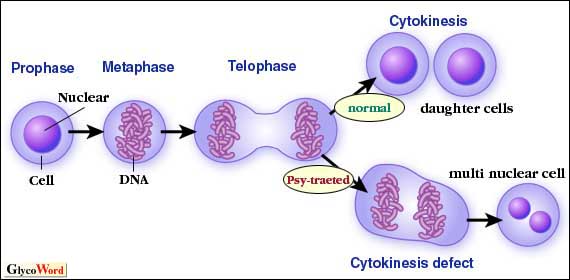
When U937 cells were incubated with Psy, multinuclear globular giant cells similar to the globoid cells in GLD were formed. During the multinuclear formation, nuclear division proceeded without cytokinesis (Figure 2). The DNA contents of these giant cells increased to 64N corresponding to 16-32 nuclei by long-term incubation with Psy. Time lapse microscopy demonstrated that the cleavage furrows formed once and disappeared during cytokinesis in the Psy-treated U937 cells. In order to study the detailed mechanism, the localization of actin, which is necessary for cell division in eukaryotes was investigated. Psy-treated U937 cells showed giant actin clots along the plasma membrane, and these clots were observed even in mitosis. Electron microscopy showed heavy vesicle accumulation in the clot area. Furthermore, filamentous structures were observed among the vesicles. The vesicles are believed to be derived from endosomes and the filamentous structures are believed to be actin filaments.
These results suggest that Psy affects vesicle transport and/or actin reorganization, resulting the abnormal cytokinesis. Although some results have been accumulated, the regulation of cytokinesis is not fully understood. A study of the Psy-induced cytokinesis inhibition would contribute not only to the effective therapy of GLD, but also to the regulation of cytokinesis. |
|||||||||||
 |
|||||||||||
| Fig. 2 Psy-treated cells proceed with nuclear division and cleavage furrow formation. However, the cells fail to complete cytokinesis and become multinuclear cells. |
|||||||||||
| Takayuki Kanazawa (Department of Signal Transductions, Graduate School of Biostudies, Kyoto University) |
|||||||||||
|
|||||||||||
| Oct. 1, 2002 | |||||||||||
|
|
|||||||||||
|
|||||||||||