



Mice Deficient in Heparan Sulfate/Heparin Biosynthetic Enzymes | ||||||||||||||||||||||||||||||||||||||||||||||||||||||||||||||||||||||||||||||
 |
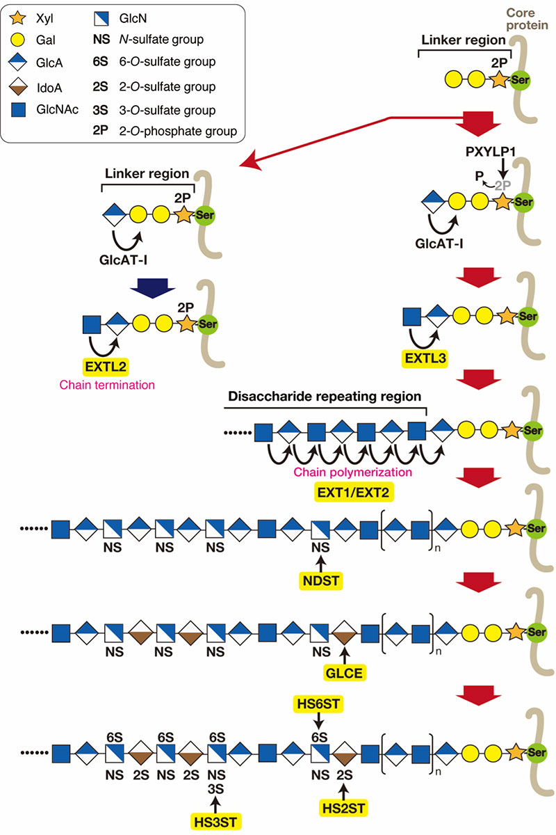
Heparan sulfate proteoglycans (HS-PGs) on cell surfaces, basement membranes, and the extracellular matrix influence biological processes by interacting with many physiologically important proteins. Thus, mice in which the genes important for HS biosynthesis are knocked out exhibit various phenotypes, including chondrodysplasia, kidney agenesis, abnormal mast cells, and lung dysfunction. On this page, some mouse models in which genes encoding HS biosynthetic enzymes are knocked out are shown.
Although HS3ST1 has long been thought to be involved in the formation of the glucosamine 3-O-sulfate structure, which is essential for the binding of heparin to the anticoagulant protein antithrombin, coagulation is normal in Hs3st1-null mice (1,2,57). Recently, it has been demonstrated that HS3ST mediates anti-inflammatory activity via antithrombin in a lipopolysaccharide-induced acute septic shock model (2). Most Hs6st1-null mice die during the late embryonic stage, and mice that survive exhibit developmental abnormalities (1,2,58-60). The 6-O-sulfation of GlcNS residues by HS6ST is critical for regulating fibroblast growth factor-dependent signaling. Consequently, it has been suggested that Hs6st1-null mice exhibit abnormal axon patterning in retinal ganglion cells, cranial axon guidance, and corpus callosum development (2,58-60). Hs6st2-deficient mice develop normally but exhibit age-dependent weight gain, impaired glucose metabolism, and insulin resistance even when fed a standard diet (61). In fetal skin-derived mast cells from Hs6st1 and Hs6st2 double-knockout mice, 6-O-sulfation was almost completely abolished, resulting in the near-total loss of tryptase and carboxypeptidase A activity. In addition, FGF signaling was partially attenuated in fetal fibroblasts (62, 63). Satomi Nadanaka & Hiroshi Kitagawa
| |||||||||||||||||||||||||||||||||||||||||||||||||||||||||||||||||||||||||||||
| References | |
|---|---|
| (1) | Mizumoto S, Shuhei Y, Sugahara K: Human Genetic Disorders and Knockout Mice Deficient in Glycosaminoglycan. BioMed Res. Int. 2014, 495764, 2014 |
| (2) | Mashima R, Okuyama T, Ohira M: Physiology and Pathophysiology of Heparan Sulfate in Animal Models: Its Biosynthesis and Degradation. Int. J. Mol. Sci. 23(4), 1963, 2022 |
| (3) | Bishop J, Schuksz M, Esko JD: Heparan sulphate proteoglycans fine-tune mammalian physiology. Nature 446, 1030-1037, 2007 |
| (4) | Nadanaka S, Kitagawa H: EXTL2-related Glycosaminoglycan Biosynthesis and Disease. TIGG 35(203) E1-5, 2023 |
| Mice | Phenotype | Original paper | Ext1 or Ext2 (global knockout) |
Global Ext1- or Ext2-deficient mice died between embryonic days 8.5 and 14.5 due to defects in mesoderm formation and impaired elongation of the egg cylinder. Ext1 hypomorphic mutation: GlcA and GlcNAc transferase activities were reduced, resulting in shortened HS chains. Furthermore, this mutation affected the Indian hedgehog (Ihh) and parathyroid hormone-related protein (PTHrP) signaling pathways. |
5) Lin X, Wei G, Shi Z, Dryer L, Esko JD, Wells DE, Matzuk MM: Disruption of gastrulation and heparan sulfate biosynthesis in Ext1-deficient mice. Dev. Biol. 224(2), 299–311, 2000 6) Stickens D, Zak BM, Rougler N, Esko JD, Werb Z: Mice deficient in Ext2 lack heparan sulfate and develop exostoses. Development. 132(22), 5055–5068, 2005 7) Koziel L, Kunath M, Kelly OG, Vortkamp A: Ext1-dependent heparan sulfate regulates the range of Ihh signaling during endochondral ossification. Dev. Cell. 6 (6), 801–813, 2004 8) Yamada S, Busse M, Ueno M, Kelly OG, Skarnes WC, Sugahara K, and Kusche-Gullberg M: Embryonic fibroblasts with a gene trap mutation in Ext1 produce short heparan sulfate chains. J. Biol. Chem. 279(31), 32134–32141, 2004 |
Ext1 (neuron-specific knockout) |
Neuron-specific Ext1-knockout mice died within 1 day after birth. In addition, dysregulation of signaling pathways, including Fgf8 and Netrin-1, led to hypoplasia in the olfactory bulb and midbrain-hindbrain regions, as well as defects in axon guidance. | 9) Inatani M, Irie F, Plump AS, Tessier-Lavigne M, Yamaguchi Y: Mammalian brain morphogenesis and midline axon guidance require heparan sulfate. Science 302(5647), 1044–1046, 2003 10) Matsumoto Y, Irie F, Inatani M. Tessier-Lavigne M, Yamaguchi Y: Netrin-1/DCC signaling in commissural axon guidance requires cell-autonomous expression of heparan sulfate. J. Neurosci. 27(6), 4342–4350, 2007 11) Yamaguchi Y, Inatani M, Matsumoto Y, Ogawa J, Irie F: Roles of heparan sulfate in mammalian brain development: current views based on the findings from ext1 conditional knockout studies. Prog. Mol. Biol. Transl. Sci. 93, 133–152, 2010s |
Ext1 (postnatal neuron-specific knockout) | No gross abnormalities in brain structure or neuronal organization were observed at the macroscopic or microscopic level. These mice exhibited autism-like behaviors; specifically, in behavioral assays, they displayed characteristics typical of autism spectrum disorder, such as reduced social interaction and repetitive behaviors. | 12) Irie F, Badie-Mahdavi H, Yamaguchi Y: Autism-like socio-communicative deficits and stereotypies in mice lacking heparan sulfate. Proc. Natl. Acad. Sci. USA 109(13), 5052–5056, 2012 | Ext1 (chondrocyte- and limb bud-specific knockout) Ext2 heterozygous mice Ext1+/−/Ext2+/− compound heterozygous mice |
These mice exhibited skeletal abnormalities associated with defective chondrocyte differentiation and maturation. These defects were similar to those observed in hereditary multiple exostoses (HME), an autosomal dominant disorder in humans. | 13) Zak BM, Schuksz M, Koyama E, Mundy C, Wells D E, Yamaguchi Y, Pacifici M, and Esko JD: Compound heterozygous loss of Ext1 and Ext2 is sufficient for formation of multiple exostoses in mouse ribs and long bones. Bone 48(5), 979–987, 2011 14) Matsumoto Y, Matsumoto K, Irie F, Fukushi JI, Stallcup WB, and Yamaguchi Y: Conditional ablation of the heparan sulfate-synthesizing enzyme Ext1 leads to dysregulation of bone morphogenic protein signaling and severe skeletal defects. J. Biol. Chem. 285(25), 19227–19234, 2010 15) Mundy C, Yasuda T, Kinumatsu T, Yamaguchi Y, Iwamoto M, Enomoto-Iwamoto M, Koyama E, Pacifici M: Synovial joint formation requires local Ext1 expression and heparan sulfate production in developing mouse embryo limbs and spine. Dev. Biol. 351(1), 70–81, 2011 16) Huegel J, Mundy C, Sgariglia F, Nygren P, Billings PC, Yamaguchi Y, Koyama E, and Pacifici M: Perichondrium phenotype and border function are regulated by Ext1 and heparan sulfate in developing long bones: a mechanism likely deranged in Hereditary Multiple Exostoses. Dev. Biol. 377(1), 100–112, 2013 17) Sgariglia F, Candela ME, and Huegel J: Epiphyseal abnormalities, trabecular bone loss and articular chondrocyte hypertrophy develop in the long bones of postnatal Ext1-deficient mice. Bone 57(1) 220–231, 2013 |
Ext1 (podocyte-specific) |
Kidney podocytes are rich in heparan sulfate proteoglycans (HSPGs), which are essential for maintaining the normal structure and signaling of the filtration barrier. Deficiency of Ext1 in podocytes leads to defective synthesis of HS chains, resulting in morphological abnormalities of the podocytes. | 18) Chen S, Wassenhove-McCarthy DJ, Yamaguchi Y, Holzman LB, Van Kuppevelt TH, Jenniskens GJ, Wijnhoven TJ, Woods AC, and McCarthy K J: Loss of heparan sulfate glycosaminoglycan assembly in podocytes does not lead to proteinuria. Kidney Int. 74(3), 289–299, 2008 | Ext1 (High endothelial venule (HEV)-specific) |
Lymphocyte homing (migration) to peripheral lymph nodes was reduced, and the contact hypersensitivity response (delayed-type hypersensitivity reaction) was impaired. | 19) Bao X, Moseman EA, Saito H, Petryanik B, Thiriot A, Hatakeyama S, Ito Y, Kawashima H, Yamaguchi Y, Lowe JB, von Andrian UH, and Fukuda M: Endothelial heparan sulfate controls chemokine presentation in recruitment of lymphocytes and dendritic cells to lymph nodes. Immunity 3(5), 817–829, 2010 20) Tsuboi K, Hirakawa J, Seki E, Imai Y, Yamaguchi Y, Fukuda M, and Kawashima H: Role of high endothelial venule-expressed heparan sulfate in chemokine presentation and lymphocyte homing. J. Immunol. 191(1), 448–455, 2013 |
Extl2 (global knockout) |
Extl2-knockout mice developed normally and produced more GAG chains than wild-type mice. Liver regeneration following carbon tetrachloride-induced liver injury was impaired in Extl2-knockout mice due to a reduced response to hepatocyte growth factor (HGF). When the STAM mouse model of non-alcoholic steatohepatitis-hepatocellular carcinoma (NASH-HCC) was applied to Extl2-knockout mice, the GAGs synthesized under Extl2-deficient conditions activated TLR4 signaling, thereby promoting the development of hepatocellular carcinoma. Furthermore, it has been demonstrated using fibroblasts that excessive GAG production resulting from Extl2 deficiency attenuates FGF2 signaling. Following spinal cord demyelinating injury in Extl2-knockout mice, excessive synthesis of proteoglycans led to the accumulation of macrophages at the lesion site. These macrophages overproduced TNFα and MMPs, which exacerbated inflammation and tissue destruction, thereby promoting axonal damage and demyelination. Aortic overexpression of GAGs exacerbates vascular calcification in a chronic kidney disease model of Extl2-knockout mice. |
21) Nadanaka S, Zhou S, Kagiyama S, Shoji N, Sugahara K, Sugihara K, Asano M, Kitagawa H: EXTL2, a member of the EXT family of tumor suppressors, controls.glycosaminoglycan biosynthesis in a xylose kinase-dependent manner. J. Biol. Chem. 288(13), 9321–9333, 2013 22) Nadanaka S, Kagiyama S, Kitagawa H: Roles of EXTL2, a member of the EXT family of tumour suppressors, in liver injury and regeneration processes. Biochem. J. 454(1), 133–145, 2013 23) Nadanaka S, Hashiguchi S, Kitagawa H: Aberrant glycosaminoglycan biosynthesis by tumor suppressor EXTL2 deficiency promotes liver inflammation and tumorigenesis through Toll-like 4 receptor signaling. FASEB J. 34(6), 8385-8401, 2020 24) Nadanaka S, Kitagawa H: Exostosin-like 2 regulates FGF2 signaling by controlling the endocytosis of FGF2. Biochem. Biophys. Acta. Gen. Subj. 1862(4), 791-799, 2018 25) Pu A, Mishra MK, Dong Y, Ghorbanigazar S, Stephenson EL, Rawji KS, Silva C, Kitagawa H, Sawcer S, Yong VW: The glycosyltransferase EXTL2 promotes proteoglycan deposition and injurious neuroinflammation following demyelination. J. Neuroinflammation 17, 220, 2020 26) Purnomo E, Emoto N, Nugrahaningsih DAA, Nakayama K, Yagi K, Heiden S, Nadanaka S, Kitagawa H, Hirata K: Glycosaminoglycan overproduction in the aorta increases aortic calcification in murine chronic kidney disease. J. Am. Heart Assoc. 2(5), e000405, 2013 |
Extl3 (global knockout and pancreatic β-cell-specific knockout) |
Global knockout: Embryonic lethal. Pancreatic β-cell-specific knockout: Resulted in morphological abnormalities of the islets and impaired proliferation of pancreatic β-cells, leading to deficient insulin secretion. |
27) Takahashi I, Noguchi N, Nata K, Yamada S, Kaneiwa T, Mizumoto S, Ikeda T, Sugihara K, Asano M, Yoshikawa T, Yamauchi A, Shervani NJ, Uruno A, Kato I, Unno M, Sugahara K, Takasawa S, Okamoto H, Sugawara A: Important role of heparan sulfate in postnatal islet growth and insulin secretion. Biochem. Biophys. Res. Commun. 383(1), 113–118, 2009 | Ndst1 (global knockout) |
Ndst1-knockout mice died postnatally and exhibited brain malformation, axon guidance defects, abnormalities in the eye and olfactory bulb, and deficient milk production due to impaired development of the lobuloalveolar structure in the mammary glands, as well as morphological abnormalities in podocytes. | 28) Fan G, Xiao L, Cheng L, Wang X, Sun B, Hu G: Targeted disruption of NDST-1 gene leads to pulmonary hypoplasia and neonatal respiratory distress in mice. FEBS Let. 467(1), 7–11, 2000 29) Ringvall M, Ledin J, Holmborn K, Van Kuppevelt T, Ellin F, Eriksson I, Olofsson A-M, Kjellén L, Forsberg E: Defective heparan sulfate biosynthesis and neonatal lethality in mice lacking N-deacetylase/N-sulfotransferase-1. J. Biol. Chem. 275(34), 25926–25930, 2000 30) Grobe K, Inatani M, Pallerla SR, Castagnola J, Yamaguchi Y, and Esko JD: Cerebral hypoplasia and craniofacial defects in mice lacking heparan sulfate Ndst1 gene function. Development. 132(16), 3777–3786, 2005 31) Pan Y, Woodbury A, Esko JD, Grobe K, Zhang X: Heparan sulfate biosynthetic gene Ndst1 is required for FGF signaling in early lens development. Development, 133(24), 4933–4944, 2006 32) Crawford BE, Garner OB, Bishop JR, Zhang D Y, Bush KT, Nigam SK, and Esko JD: Loss of the heparan sulfate sulfotransferase, Ndst1, in mammary epithelial cells selectively blocks lobuloalveolar development in mice. PLoS ONE, 5(5), e10691, 2010 33) Garner OB, Bush KT, Nigam KB, Yamaguchi Y, Xu D, Esko JD, Nigam SK: Stage-dependent regulation of mammary ductal branching by heparan sulfate and HGF-cMet signaling. Dev. Biol. 355(2), 394–403, 2011 34) Axelsson J, Xu D, Kang BN, Nussbacher JK, Handel T M, Ley K, Sriramarao P, Esko JD: Inactivation of heparan sulfate 2-O-sulfotransferase accentuates neutrophil infiltration during acute inflammation in mice. Blood 120(8), 1742–1751, 2012 35) Sugar T, Wassenhove-McCarthy DJ, Esko JD, van Kuppevelt TH, Holzman L, McCarthy KJ: Podocyte-specific deletion of NDST1, a key enzyme in the sulfation of heparan sulfate glycosaminoglycans, leads to abnormalities in podocyte organization in vivo. Kidney Int. 85(2), 307-318, 2013 |
Ndst1 (liver-specific knockout) |
Accumulation of triglyceride-rich lipoprotein particles was observed, resulting from the impaired clearance of cholesterol-rich lipoprotein particles. | 36) MacArthur JM, Bishop JR, Stanford KI, Wang L, Bensadoun A, Witztum JL, Esko JD, Liver heparan sulfate proteoglycans mediate clearance of triglyceride-rich lipoproteins independently of LDL receptor family members. J. Clin. Invest. 117(1), 53–164, 2007 37) Foley EM, Gordts PLSM, Stanford KI, Gonzales J C, Lawrence R, Stoddard N, Esko JD, Hepatic remnant lipoprotein clearance by heparan sulfate proteoglycans and low-density lipoprotein receptors depend on dietary conditions in mice. Arterioscler. Thromb.Vasc. Biol. 33(9), 2065–2074, 2013 |
Ndst1 (endothelial cell-specific knockout) |
Experimental tumor growth and angiogenesis were suppressed, including reduced tumor microvessel density and tumor-associated branching, due to altered responses to fibroblast growth factor 2 (FGF2) and vascular endothelial growth factor (VEGF). Attenuation of allergic airway inflammation was also observed. | 38) Fuster MM, Wang L, Castagnola J, Sikora L, Reddi K, Lee PHA, Radek KA, Schuksz M, Bishop JR, Gallo RL, Sriramarao P, Esko JD: Genetic alteration of endothelial heparan sulfate selectively inhibits tumor angiogenesis. J. Cell Biol. 177(3), 539–549, 2007 39) Zuberi RI, Ge XN, Jiang S, Bahaie NS, Kang BN, Hosseinkhani RM, Frenzel EM, Fuster MM, Esko JD, Rao SP, Sriramarao P: Deficiency of endothelial heparan sulfates attenuates allergic airway inflammation. J. Immunol. 183(6), 3971–3979, 2009 |
Ndst2 (global knockout) |
Ndst2-deficient mice are viable and fertile, but they fail to synthesize heparin in mast cells. Consequently, this leads to morphological changes in granules and a marked reduction in the content of stored proteases. | 40) Humphries DE, Wong GW, Friend DS, Gurish MF, Qiu W-T, Huang C, Sharpe AH, Stevens RL: Heparin is essential for the storage of specific granule proteases in mast cells. Nature 400(6746), 769–772, 1999 41) Forsberg E, Pejler G, Ringvall M, Lunderius C, Tomasini-Johansson B, Kusche-Gullberg M, Eriksson I, Ledin J, Hellman L, Kjellén L.: Abnormal mast cells in mice deficient in a heparin-synthesizing enzyme. Nature 400(6746), 773–776, 1999 42) Karlsen TV, Iversen VV, Forsberg E, Kjellén L, Reed R K, Gjerde E-A B: Neurogenic inflammation in mice deficient in heparin-synthesizing enzyme. Am. J. Physiol. 286(3), H884–H888, 2004 |
Ndst3 (global knockout) |
Ndst3-deficient mice develop and breed normally, exhibiting only subtle hematological and behavioral abnormalities due to a minor reduction in HS sulfation. Analysis of the adult brain revealed that NDST3 contributes to HS sulfation in specific brain regions. Ndst2;Ndst3 double-deficient mice also develop normally, indicating that both isoforms are not essential for development. In contrast, Ndst1;Ndst3 double-deficient mice display severe HS undersulfation and associated developmental defects. NDST3 plays a critical role in HS synthesis in the absence of NDST1. |
43) Pallerla SR, Lawrence R, Lewejohann L, Pan Y, Fischer T, Schlomann U, Zhang X, Esko JD, Grobe K: Altered heparan sulfate structure in mice with deleted NDST3 gene function. J. Biol. Chem. 283(24), 16885–16894, 2008 | Glce (global knockout) |
Glce-deficient mice died shortly after birth and exhibited renal agenesis (dysplasia), shortened body length, and lung defects, as well as developmental abnormalities in lymphoid organs, including the spleen, thymus, and lymph nodes. The impairment in early thymic development resulted from disrupted signaling of fibroblast growth factor (FGF) 2, FGF10, and bone morphogenetic protein 4 (BMP4). To investigate B-cell development and differentiation, fetal liver hematopoietic stem cells from Glce-/- mice were transplanted into lymphocyte-deficient Rag2-/-γc-/- mice. This study demonstrated that Glce deficiency impaired B-cell maturation, resulting in reduced plasma cell numbers and immunoglobulin levels. Since Glce-mediated modification of HS chains is critical for the binding of a proliferation-inducing ligand (APRIL), Glce-deficient plasma cells were unable to respond to APRIL-mediated survival signals. |
44) Li J-P, Gong F, Hagner-McWhirter Å, Forsberg E, Åbrink M, Kisilevsky R, Zhang X, Lindahl U: Targeted disruption of a murine glucuronyl C5-epimerase gene results in heparan sulfate lacking L-iduronic acid and in neonatal lethality. J. Biol. Chem. 278(31), 28363–28366, 2003 45) Jia J, Maccarana M, Zhang X, Bespalov M, Lindahl U, Li J-P: Lack of L-iduronic acid in heparan sulfate affects interaction with growth factors and cell signaling. J. Biol. Chem. 284(23), 15942–15950, 2009 46) Reijmers R. M., Vondenhoff M. F. R., Roozendaal R., Kuil A., Li J.-P., Spaargaren M., Pals S. T., and Mebius R. E., Impaired lymphoid organ development in mice lacking the heparan sulfate modifying enzyme glucuronyl C5-epimerase. J. Immunol. 184(7), 2010 47) Reijmers R. M., Groen R. W. J., Kuil A., Weijer K., Kimberley F. C., Medema J. P., Van Kuppevelt T. H., Li J.-P., Spaargaren M., and Pals S. T., Disruption of heparan sulfate proteoglycan conformation perturbs B-cell maturation and APRIL-mediated plasma cell survival. Blood. 117(23), 6162–6171, 2011 |
Hs2st (global knockout) |
Hs2st-deficient mice died shortly after birth and exhibited renal agenesis, as well as defects in the eye, skeleton, and retinal axon guidance. | 48) Bullock SL, Fletcher JM, Beddington RSP, Wilson VA: Renal agenesis in mice homozygous for a gene trap mutation in the gene encoding heparan sulfate 2-sulfotransferase. Genes Dev. 12(12), 1894–1906, 1998 49) Shah MM, Sakurai H, Sweeney DE, Gallegos TF, Bush KT, Esko JD, Nigam SK: Hs2st mediated kidney mesenchyme induction regulates early ureteric bud branching. Dev. Biol. 339(2), 354–365, 2010 50) Merry CLR, Bullock SL, Swan DC, Backen AC, Lyon M, Beddington RSP, Wilson VA, Gallagher JT, The Molecular Phenotype of Heparan Sulfate in the Hs2st-/- Mutant Mouse. J. Biol. Chem. 276(38), 35429–35434, 2001 51) McLaughlin D, Karlsson F, Tian N, Pratt T, Bullock SL, Wilson VA, Price DJ, Mason JO: Specific modification of heparan sulphate is required for normal cerebral cortical development. Mech. Dev. 120(12) 1481–1488, 2003 52) Pratt T, Conway CD, Tian NM M-L, Price DJ, Mason JO: Heparan sulphation patterns generated by specific heparan sulfotransferase enzymes direct distinct aspects of retinal axon guidance at the optic chiasm. J. Neurosci. 26(26), 6911–6923, 2006 53) Conway CD, Howe KM, Nettleton NK, Price DJ, Mason JO, Pratt T: Heparan sulfate sugar modifications mediate the functions of Slits and other factors needed for mouse forebrain commissure development. J. Neurosci. 31(6), 1955–1970, 2011 54) Conway CD, Price DJ, Pratt T. Mason JO, Analysis of axon guidance defects at the optic chiasm in heparan sulphate sulphotransferase compound mutant mice. J. Anatomy 219(6), 734–742, 2011 |
Hs2st (endothelial cell-specific and myeloid cell-specific knockout) |
Increased binding to IL-8 and macrophage inflammatory protein-2 (MIP-2) promoted neutrophil infiltration. | 55) Axelsson J, Xu D, Kang BN, Nussbacher JK, Handel T M, Ley K, Sriramarao P, Esko JD: Inactivation of heparan sulfate 2-O-sulfotransferase accentuates neutrophil infiltration during acute inflammation in mice. Blood. 120(8) 1742–1751, 2012 | Hs2st (liver-specific knockout) |
Plasma triglycerides accumulated, and the uptake of very-low-density lipoprotein (VLDL) was reduced. | 56) Stanford KI, Wang L, Castagnola J, Song D, Bishop JR, Brown JR. Lawrence R, Bai X, Habuchi H, Tanaka M, Cardoso WV, Kimata K, Esko JD: Heparan sulfate 2-O-sulfotransferase is required for triglyceride-rich lipoprotein clearance. J. Biol. Chem. 285(1), 286–294, 2010 | Hs3st1 (global knockout) |
Hs3st1-deficient mice developed normally and exhibited normal anticoagulant activity. Since the glucosamine (GlcN) 3-O-sulfated structure is essential for the anticoagulant activity of heparin and HS, other members of the HS3ST family may compensate for the loss of HS3ST1. | 57) HajMohammadi S, Enjyoji K, Princivalle M, Christi P, Lech M, Beeler D, Rayburn H, Schwartz JJ, Barzegar S, De Agostini AI, Post MJ, Rosenberg RD, Shworak N. W: Normal levels of anticoagulant heparan sulfate are not essential for normal hemostasis. J. Clin. Invest. 111(7), 989–999, 2003 | Hs6st1 (global knockout) |
Hs6st1-deficient mice died in the late embryonic stage, were smaller than wild-type mice at birth, and exhibited defects in retinal axon guidance due to impaired Slit-Robo signaling. | 58) Pratt T, Conway CD, Tian NM M-L, Price DJ, Mason JO: Heparan sulphation patterns generated by specific heparan sulfotransferase enzymes direct distinct aspects of retinal axon guidance at the optic chiasm. J. Neurosci. 26(26), 6911–6923, 2006 59) Conway CD, Howe KM, Nettleton NK, Price DJ, Mason JO, Pratt T: Heparan sulfate sugar modifications mediate the functions of Slits and other factors needed for mouse forebrain commissure development. J. Neurosci. 31(6), 1955–1970, 2011 60) Habuchi H, Nagai N, Sugaya N, Atsumi F, Stevens RL, Kimata K: Mice deficient in heparan sulfate 6-O-sulfotransferase-1 exhibit defective heparan sulfate biosynthesis, abnormal placentation, and late embryonic lethality. J. Biol. Chem. 282(21), 15578–15588, 2007 |
Hs6st2 (global knockout) |
Hs6st2-deficient mice develop normally, but male mice fed a standard diet exhibit age-dependent weight gain, glucose intolerance, and insulin resistance. | 61) Nagai N, Habuchi H, Sugaya N, Nakamura M, Imamura T, Watanabe H, Kimata K: Involvement of heparan sulfate 6-O-sulfation in the regulation of energy metabolism and the alteration of thyroid hormone levels in male mice. Glycobiology 23(8), 980–992, 2013 | Hs6st1;Hs6st2 (global double-knockout) |
In fetal skin-derived mast cells, 6-O-sulfation of heparin was almost completely abolished, and the activities of proteases such as tryptase and carboxypeptidase A were markedly reduced, accompanied by abnormal storage. In contrast, the activity and storage of chymase were maintained. In fetal fibroblasts, FGF signaling was partially attenuated. | 62) Anower-E-Khuda MF, Habuchi H, Nagai N, Habuchi O, Yokochi T, Kimata K: Heparan sulfate 6-O-sulfotransferase isoform-dependent regulatory effects of heparin on the activities of various proteases in mast cells and the biosynthesis of 6-O-sulfated heparin. J. Biol. Chem. 288(6), 3705–3717, 2013 63) Sugaya N, Habuchi H, Nagai N, Ashikari-Hada S, Kimata K: 6-O-sulfation of heparan sulfate differentially regulates various fibroblast growth factor-dependent signalings in culture. J. Biol. Chem. 283(16), 10366–10376, 2008 |
|---|
Jul. 10, 2026