
Hideo Mochizuki
https://orcid.org/0009-0003-1051-630X
Hideo Mochizuki graduated from the Faculty of Science, Konan University in 1983 and obtained his Ph.D. from the Graduate School, Kanazawa University in 1990. He joined Seikagaku Corporation in 1990 and left the company in 2025. From 2002 to 2004, he served as a Visiting Researcher at the Institute for Molecular Science of Medicine, Aichi Medical University, directed by his lifelong master Professor Koji Kimata. Since demonstrating the presence of the tetrasulfated disaccharide unit in heparan sulfate in 2003, he has continued to work in this field for over 20 years.
Heparan sulfate (HS) is a long linear glyco-chain component of proteoglycans on cell surfaces and within the extracellular matrix in most animal species, ranging from hydra to humans1. HS regulates various physiological processes by interaction with numerous proteins including growth factors, morphogens, cytokines, receptors, enzymes, and extracellular matrix proteins2. Additionally, some pathogens exploit HS on the host cell surface as a receptor for invasion3. HS has a complex structure consisting of a mixture of regions with high-density, heterogeneous sulfation and regions with almost no sulfate groups. The binding specificity of HS for each functional protein is thought to be dependent on the specific arrangement of sulfate groups within the sulfated regions4.
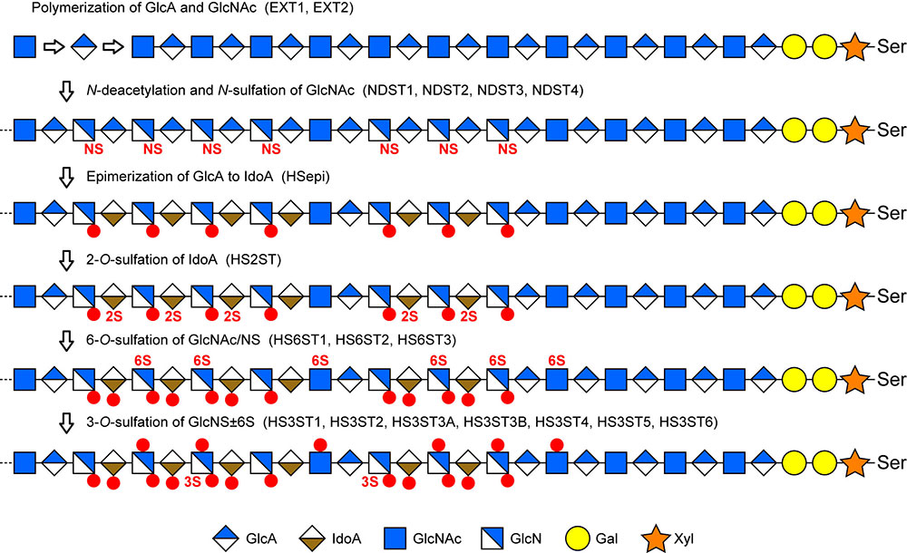
The capacity of cells to produce HS with such a variety of modifications depends on the mechanism of HS biosynthesis. The biosynthesis occurs via the reaction pathway shown in Figure 1, catalyzed by enzymes localized in the Golgi apparatus. It is noteworthy that some of the reactions within this pathway are catalyzed by multiple isoforms, as indicated in parentheses. These isoforms exhibit varying specificities toward the structures surrounding the sulfation site. The biosynthesis begins with the formation of a repeating disaccharide structure, -GlcA-β1,4-GlcNAc-α1,4-, by alternate addition of N-acetylglucosamine (GlcNAc) and glucuronic acid (GlcA) residues to the tetrasaccharide of GlcA-Gal-Gal-Xyl- bound to the serine residue of the core protein. The repeating disaccharide units range in length from several dozen to over 200 units. Subsequently, part of this linear chain undergoes N-deacetylation and N-sulfation, thereby forming the basis for the sulfated region. Next, GlcA residues undergo isomerization to form iduronic acid (IdoA). The IdoA, rarely GlcA, residues undergo 2-O-sulfation, and glucosamine residues undergo 6-O-sulfation. In addition, a rare yet functionally significant modification, 3-O-sulfation of the glucosamine residues, also occurs. It is believed that the HS biosynthetic enzymes form a complex, known as GAGosome, within the Golgi apparatus, and that interactions between these enzymes regulate tailored biosynthesis. The biosynthesis yields 12 different sulfation patterns of disaccharide units. In sulfated regions where these units are arranged in a linear sequence, an enormous number of combinations becomes possible, thereby enabling the formation of binding structures for individual functional proteins. Therefore, rare disaccharide units are considered key components in the formation of specific binding structures.
From the above perspective, a significant area of research is the elucidation of the functions of the 3-O-sulfated (3S) structure5. Although disaccharide units with the 3S structure are usually less than 2% of the total disaccharides, mammals, including humans, harbor 7 isoform genes for the enzyme that catalyzes this rare modification reaction, namely heparan sulfate 3-O-sulfotransferase (HS3ST). The presence of multiple isoforms with varying specificities is thought to be required for the recognition of diverse surrounding structures and the 3-O-sulfation of specific glucosamine residues. Figure 2 shows the amino acid alignment of HS3ST isoforms. The C-terminal region of HS3ST forms a globular domain containing the catalytic site and exhibits high amino acid sequence homology across all isoforms (blue letters). In contrast, the N-terminal region, known as the stem region, shows no homology whatsoever. The stem regions contain a transmembrane segment (orange letters) that anchors the enzyme to the Golgi membrane. Only HS3ST1 lacks this segment and localizes to the Golgi by interacting with other proteins. The function of stem regions remains unclear.
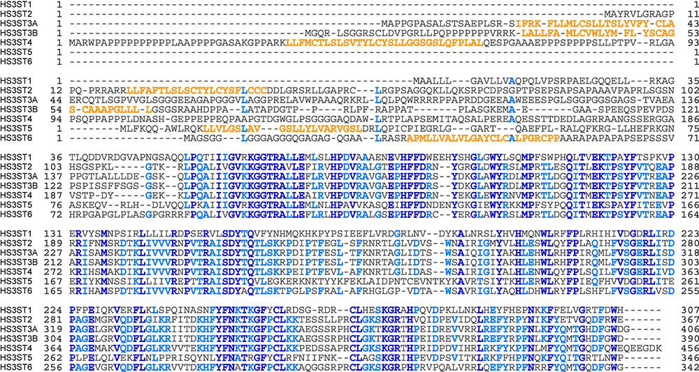
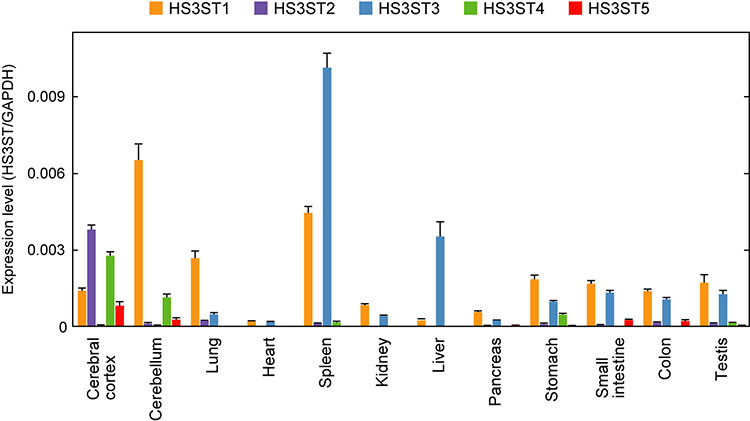
Figure 3 shows the expression levels of HS3ST isoforms in human organs. The expression patterns of isoforms vary significantly across organs, suggesting differences in the 3S structures required for each organ's function. Particularly in the brain, HS3ST2, HS3ST4, and HS3ST5 are specifically expressed, and the 3S structures formed by these isoforms may regulate neural function.
Research on the 3S structure has been in progress for a long time, with origins dating back to 1980 when Lindahl et al. first identified the 3S glucosamine residue within the antithrombin (AT)-binding structure of heparin6. Heparin is a highly sulfated form of HS synthesized by mast cells. Due to its medical significance, the AT-binding 3S structure has been thoroughly investigated. The pentasaccharide shown in Figure 4 has the minimum structure necessary for AT-binding, and its binding ability is lost upon removal of the central 3-O-sulfate group7. The AT-binding 3S structure also represents the sole demonstration of a clear structure-function relationship8. The enzyme responsible for synthesizing the AT-binding structure (HS3ST1) has been purified and cloned as a first enzyme of the HS3ST gene family9,10. Whereas HS3ST1 was identified on the basis of physiological function, the other six isoforms were identified in the DNA database as genes homologous with the HS3ST1 gene11-13. All these genes have been cloned, and the enzymatic properties have been investigated in vitro. Interestingly, unlike HS3ST1, five of the isoforms (HS3ST2, HS3ST3A, HS3ST3B, HS3ST4, and HS3ST6) do not form the AT-binding 3S structure. HS3ST5 forms both AT-binding and non-AT-binding 3S structures. Although the 3S structure is expected to play a crucial role in various physiological functions of HS, reports on its physiological activities other than anticoagulation are limited. The analysis of rare 3S structures in biological samples poses a significant challenge, hindering progress in this research field. Nevertheless, analyses of HS3ST gene expression and studies using synthetic 3S HS suggest that the 3S structure plays a crucial role in multiple physiological processes. For instance, HS sulfated by HS3ST2 activates the semaphorin-induced growth cone collapse14. In the nervous system of Caenorhabditis elegans, the HS3ST gene has been demonstrated to regulate the branching of neurites15. Inhibiting the expression of HS3STB gene in Drosophila melanogaster results in abnormalities in the Notch signaling pathway16. Expression of the HS3ST3 gene is necessary for the morphogenesis of mouse salivary glands17. HS3ST3A/HS3ST3B double-knockout mice reportedly exhibit a significantly elevated incidence of ureter duplication18. In the pineal gland of rats, which controls the circadian rhythm, HS3ST2 has been shown to be expressed only during the daytime19.

Heparin lyases have a long history of use in compositional analyses of HS. Three types of HS degradation enzymes with different specificity to the sulfated structure, named heparin lyase I, II and III, have been isolated from Pedobacter heparinus or Bacteroides eggerthii, and are commercially available. Using a mixture of three lyases, HS can be digested almost completely into unsaturated (∆) disaccharides, with the exception of the AT-binding 3S structure. The glucosaminidic linkage adjacent to GlcA-GlcNS3S±6S, which plays a critical role in AT-binding activity, is resistant to digestion, resulting in the generation of ∆tetrasaccharides20. The other 3S structures are not resistant to lyase I and are thus digested to ∆disaccharides21. In the digestion products, a double bond between C4 and C5 is introduced into the non-reducing terminal GlcA or IdoA residue to form 4,5-unsaturated uronic acid (∆UA). Figure 5 shows the 13 ∆saccharides produced by the lyase reaction. The structural composition of HS can be determined by separating and quantifying these ∆saccharides in the digestion products using high performance liquid chromatography (HPLC). This method, known as disaccharide analysis, has been widely used for many years, but it was limited to measuring only eight non-3S disaccharides. This was because the 3S structures present in HS, with the exception of the AT-binding structure, had not been clearly identified, and the standard 3S saccharides were unavailable.
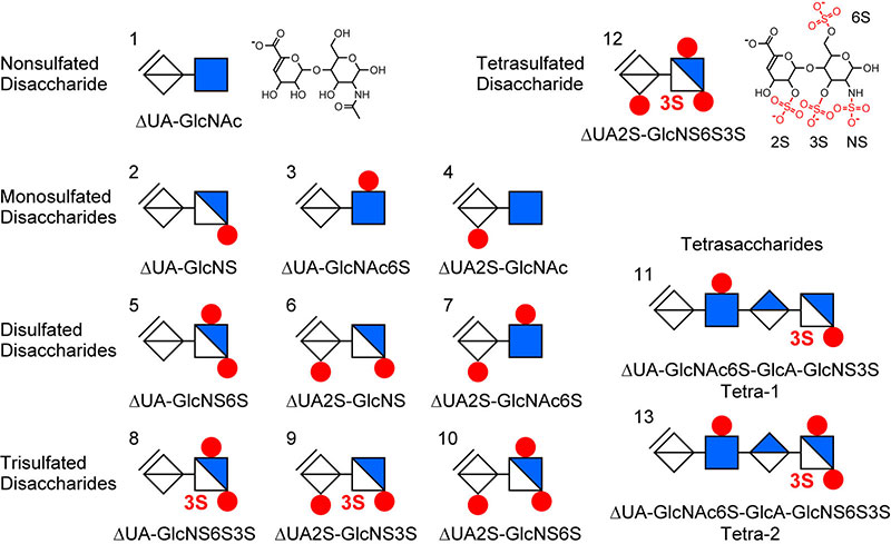
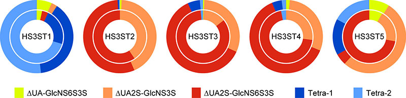
We previously analyzed the reaction products of HS3ST isoforms, and identified five 3S components as a digestion product of heparin lyases22. Of these five, three were disaccharides, including ∆UA-GlcNS6S3S, ∆UA2S-GlcNS3S, and ∆UA2S-GlcNS6S3S21. The remaining two components were lyase-resistant tetrasaccharides with structures ∆UA-GlcNAc6S-GlcA-GlcNS3S and ∆UA-GlcNAc6S-GlcA-GlcNS6S3S, named Tetra-1 and Tetra-2, respectively23. Figure 6 illustrates the production ratio of the 3S components formed by each isoform using HS (outer chart) or heparin (inner chart) as a substrate. In the case of HS3ST1, two tetrasaccharides were the principal products, whereas almost no tetrasaccharides were formed by HS3ST2, HS3ST3, or HS3ST4, and the principal products were ∆UA2S-GlcNS3S and ∆UA2S-GlcNS6S3S. It should be noted that since the catalytic domains of HS3ST3A and HS3ST3B exhibit identical amino acid sequences, their reaction specificities are also the same. Unlike other isoforms, HS3ST5 forms both disaccharides and tetrasaccharides, and in addition shows a marked difference in specificity for the two substrates. HS3ST6 had not yet been reported at the time of this examination, thus it was not measured. A subsequent report has demonstrated that HS3ST6 does not produce the AT-binding structure, and has the reaction specificity similar to that of HS3ST313.
In the next stage, we aimed to establish a method for the quantification of all 13 components, including the five 3S components24. To this end, we employed the method established by Toyoda et al., which utilizes reverse-phase ion-pair HPLC and post-column fluorescent labeling25. The method is highly sensitive and specific for detecting the reducing terminal produced by the lyase reaction, making it suitable for measuring trace components such as 3S units. The HPLC conditions were optimized to achieve the baseline separation of all 13 components. In addition, we prepared a standard HS, named HS13, composed of the 13 components in known quantities23. A standard mixture of the 13 components can be obtained by digesting HS13 with the heparin lyases (Figure 7A).
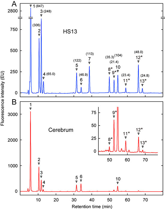
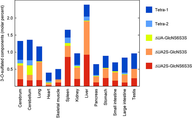
We then performed a compositional analysis of HS derived from various rat organs23. While several reports have demonstrated compositional differences of HS from various animal organs, none have made reference to the 3S structure. Our report is the first to demonstrate the overall distribution of 3S components in vivo. Figure 7B shows a representative chromatogram obtained by analyzing HS from the cerebrum. Figure 8 illustrates the distribution of five 3S components across 13 organs. Interestingly, each 3S component exhibited a distinct organ distribution, suggesting a correlation between organ-specific functions and 3S structures. However, it is important to note that organs are comprised of various types of tissues, which are in turn composed of cells with different functions. Therefore, the analysis of the 3S structure in individual tissues or cells is a future challenge that must be addressed in order to understand the 3S structure involved in each physiological function within organs.
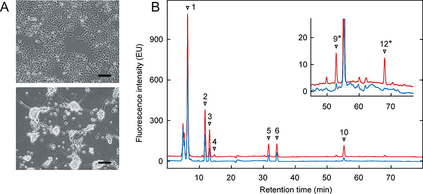
We have previously conducted an analysis of HS from cultured cells to validate the present method24. Murine embryonal carcinoma P19 cells differentiate into neurons when stimulated by retinoic acid (Figure 9A). Figure 9B shows the compositional analysis of HS derived from undifferentiated (blue chromatogram) and differentiated P19 cells (red chromatogram). Significant peaks of ∆UA2S-GlcNS3S and ∆UA2S-GlcNS6S3S were detected only in the chromatogram derived from the differentiated neurons.
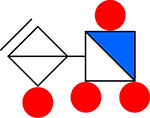
We have detected the ∆UA2S-GlcNS6S3S unit, with all four sulfation sites modified as illustrated in Figure 10, from natural HS for the first time21. In addition, we have confirmed that this unit is the major reaction product of HS3ST isoforms, which form non-AT-binding 3S structures. Although the function of this unit remains to be elucidated, we speculate that this most sulfated structure may play a specific physiological role. The liver and spleen, which contain high levels of this unit, are organs of particular interest.
Numerous studies have demonstrated that HS has the capacity to bind with at least several hundred types of proteins in a functional manner. However, research on the specific binding structures of HS has not advanced. The absence of established methodologies for analyzing the complex and heterogeneous structure of HS is hindering research progress. Moreover, the delay in research on the 3S structure is also considered a contributing factor. Due to the lack of knowledge regarding the 3S structure, much of the research to date has proceeded without considering its involvement. As a result, the structure-function relationships of HS may remain unclear. Therefore, the author believes that it is essential to re-examine the potential involvement of the 3S structure in the known physiological activities of HS.