
Kenichi Kasai
I graduated from the Department of Biophysics and Biochemistry, Faculty of Science, The University of Tokyo. After the master course, I became a researcher at the Institute of Physicochemical Biology (Paris, France), then assistant professor and associate professor in the Faculty of Pharmaceutical Sciences, Hokkaido University, and professor in the Faculty of Pharmaceutical Sciences, Teikyo University. Now I am a professor emeritus at Teikyo University. I used to study nuclease and protease, but I detoured into the field of lectin research and was captivated by glycobiology. This new field has removed the scales from my eyes so many times.
Galectin is a well-known molecular entity but remains difficult to understand. Although it appears on a wide variety of stages, it is seldom the main actor; its role seems to be supporting but indispensable. It acts with a wide variety of partners whatever their identity (protein, lipid, or proteoglycan) or position, as long as they are wearing a favorite costume (glycan). Therefore, almost all (more than 10,000) extracellular proteins can be its partners, although its relationships with them are not so intimate. This wide and shallow sociality makes the essential nature of galectin difficult to understand. With these aspects in mind, I would like to review some basic points concerning galectin and its weak interactions.
 A bird’s-eye view of galectin (Illustrated by Hiroko UCHIDA)
The ultimate fundamental basis of life resides in the interactions of biomolecules. Above all, strictly specific and strong interactions of biomolecules ensure the certainty, reliability, and reproducibility of life. This role in life’s processes is played mainly by proteins and has been understood in terms of lock-and-key relationships, such as enzyme and substrate, signal molecule and receptor, and antigen and antibody.
In reality, however, living organisms also commonly employ weak and loosely-specific interactions, including glycan recognition by lectin. Weakness and ambiguity are unlikely virtues and tend to be undervalued. Because conjugates generated by weak interaction are easily dissociable, they are difficult to detect and there is a lack of powerful research tools for analysis. Although it is often undervalued, weakly interacting couples are actually indispensable as they are closely associated with the essence of life. So, they should not be overlooked.
For example, an active protein binds not only to its main partner with which it can form a strong bond, but also often transiently to many other molecules, which have regulative roles (e.g., promotion, suppression). However, such auxiliary molecules seem to be uncapturable by the pulldown method or the like and hence are mostly overlooked because of weak binding forces.
The inside of our body is constantly monitored by a wide variety of sensors. Any sensor of change in molecule number must remove or eject the current target molecule from the binding site to prepare for the arrival of the next target molecule. If the target molecule is bound by a strong force, the sensor will be rendered inoperative because the target molecule cannot be expelled immediately. The sensor which binds weakly the target molecule is more appropriate for the purpose.
The cell membrane is comprised not only of the primary component phospholipids, but also glycolipids, which are drawn into aggregates by weak interactions, and proteins that migrate to the region to form rafts and the like. This construction serves as a center of various key activities of living organisms. Strong and specific interactions are not the only interactions worth paying attention. Weak and loose interactions support important biological tasks, without being well noticed in a wide variety of scenarios.
The major advantage of weak bonding is the absence of sustainable contact. Weakly bonded molecular species have large dissociation constants (Kd). The dissociation rate constant k-1 is relatively large because Kd = k-1/k+1; therefore, the bound state is impermanent and short lived. This is of paramount importance in biological activities.
Living organisms must quickly and accurately respond to the dynamic ever-changing conditions around them. Because many biological regulatory processes occur transiently, various systems need to be switched on and off frequently. The on-off switch is often based on mutual binding of molecules (e.g., binding between an effector molecule and protein).
Thus a system, once started, must be turned off as soon as possible after its purpose is fulfilled.
While life makes the best use of various approaches, the solution to a problem will be easiest if the conjugate dissociates soon after forming. Strong bonds take a long time to dissociate because their k-1 are usually small. Weak interactions are more suitable to a quick switching off.
In body processes such as tissue building and antigen-antibody reactions, when it is desirable to maintain conjugate integrity, strong interactions are preferred; whereas in cases of dynamic interaction phenomena, weak binding is more suitable.
Overall, weak interactions have loose specificity. There are proteins whose keyholes allow some play accommodating even somewhat structurally different molecules as partners. Lectins, for example, bind to multiple glycan units. Of course, the binding force is variable, stronger bonds do not always mean greater advantages. Even for a glycan with weak binding force, the likelihood of binding increases with increasing numbers of units. In strong interactions, specific associations are always selected. In contrast, even common glycans have chances of selection.
This may obscure the causality and affect scheduled or established approaches. However, this is not always disadvantageous. Living organisms are always subject to unexpected dangers. One-patterned responses are unlikely to overcome such abnormal situations and prevent collapse of survival chance. Availability of second and third options in emergencies is important to risk management.
Weak and loose interactions are never inferior to their counterparts. They can be surprisingly useful if their features are wisely utilized. Hydrophobic columns in common use for high-performance liquid chromatography, for example, are capable of distinctly separating dozens of molecular species (high resolution). This is due to extremely weak, almost non-specific interactions. In particular, the extremely fast binding dissociation performs several tens of thousands of checks repeatedly during column passage (through a vast theoretical number of plates), dramatically widening differences in binding force that are otherwise very small. The scope of applicable substances is wide because there is almost no specificity.
Furthermore, resolution can be increased dramatically by two-dimensional data handling and use of two columns having different characteristics.
Organisms employ the same principle to distinguish among a great many molecular entities. Olfaction is a good example. Reportedly several hundred thousand odorant molecules are sensed by a much smaller number of olfactory receptors (approximately 400 receptors in human beings). The olfactory receptors have loose specificity, binding to multiple odorant molecules. In addition, multiple receptors sense one type of odorant molecule. The brain comprehensively processes the thus-obtained data to achieve surprisingly fine distinctions. Although the specific mechanism is unknown, something like a multidimensional extension from two-dimensional analysis may be performed. Life has long used this principle to increase the resolution of the input used for recognition and much before the idea of two-dimensional analysis was introduced by analytical chemists. Moreover, weak binding force seems to be favorable for the quick recovery of receptor binding.
Hence, animals succeeded in achieving the wonderful capacity of distinguishing multiple smells using multiple sensors of relatively loose specificity. The same applies to the senses of taste and color vision.
Information glycan is essential for diplomacy between cells, including cell contact formation, information exchange, trading, tissue organization, and defense. When the first multicellular organism was born on Earth, direct and indirect interactions between cells in individuals became the top priority, and glycan was selected as the supporting material.
The cell is wrapped in a wide variety of complex glycans. Most extracellularly working proteins are decorated with glycans 1.
Although the roles of glycan are not specifically discussed here, a bird’s eye view of its significance is given below. Glycans help eliminate the uniformity of cells and proteins and make them heterogeneous. Clone members are thus individuals and diverse.
In multicellular organisms, all cells wear a coat of glycoconjugate. Because glycan biosynthesis is rather haphazard, small differences in costume design can occur among different cells of the same type, which lead to differences in affinity for lectins and other sugar-recognizing proteins.
Certain types of cell growth factors have a proteoglycan-binding site, at which they are assisted and regulated during signal transduction. In addition to having an ACE-2 binding site, the coronavirus spike has a glycan-binding site, which seems to increase coronavirus infection efficiency. As surface glycan differs minutely among different cells, signal responsiveness, pathogen resistance, and other aspects vary among cells. This diversity contributes to the flexibility and durability of living organisms, thus increasing their ability to manage risk.
Proteins are produced homogeneously like clones, with no individual differences, under strict quality control. Extracellularly, working proteins lose homogeneity upon receipt of glycan. As a result, their fates are variable. The effect on their destination, collaborators, lifespans, and other factors produce diversity in the final outcome, even in cells with the same function.
While uniformity is needed for the fundamentals of life, organisms cannot endure environmental changes, survive, and evolve without diversity. One of the important roles of glycan may be to promote the transition from uniformity to diversity.
The binding force of lectin is generally weaker than that of antibodies, its Kd being often approximately 10-4 to 10-6 M. A considerable number of biological phenomena are triggered by lectin-glycan binding. These levels of Kd seem to be suitable for control functions such as switching on and off.
In addition, lectin specificity is generally lower than the specificity of enzymes and signal molecule receptors. Although selectin has extremely high specificity, this is a somewhat exceptional case for the dispatch of leukocytes to the inflammation site.
Acting as Don Juans in the world of proteins, many lectins interact with multiple glycans. Because difference in the binding force for the glycans is not so outstanding, no particular glycan is destined to become the the first choice. Why is lectin so capricious?
This is likely attributable to the fact that glycans act as the third information medium in living organisms. Glycan is a highly problematic polymer, fabricated without design plan or synthesis apparatus, widely varying in structure with no guarantee that any of them will be as desired. The problem with the use of this troublesome mediator may have been solved compromisingly by partnering it with somewhat similar molecules rather than strictly specific molecules.
In the history of life that followed, however, evolution has not brushed up the binding characteristics of lectin. These halfway binding characteristics seem to have contributed to the diversity, flexibility, and elasticity, hence increasing the profoundness of life. As such, these characteristics are now established as useful properties.
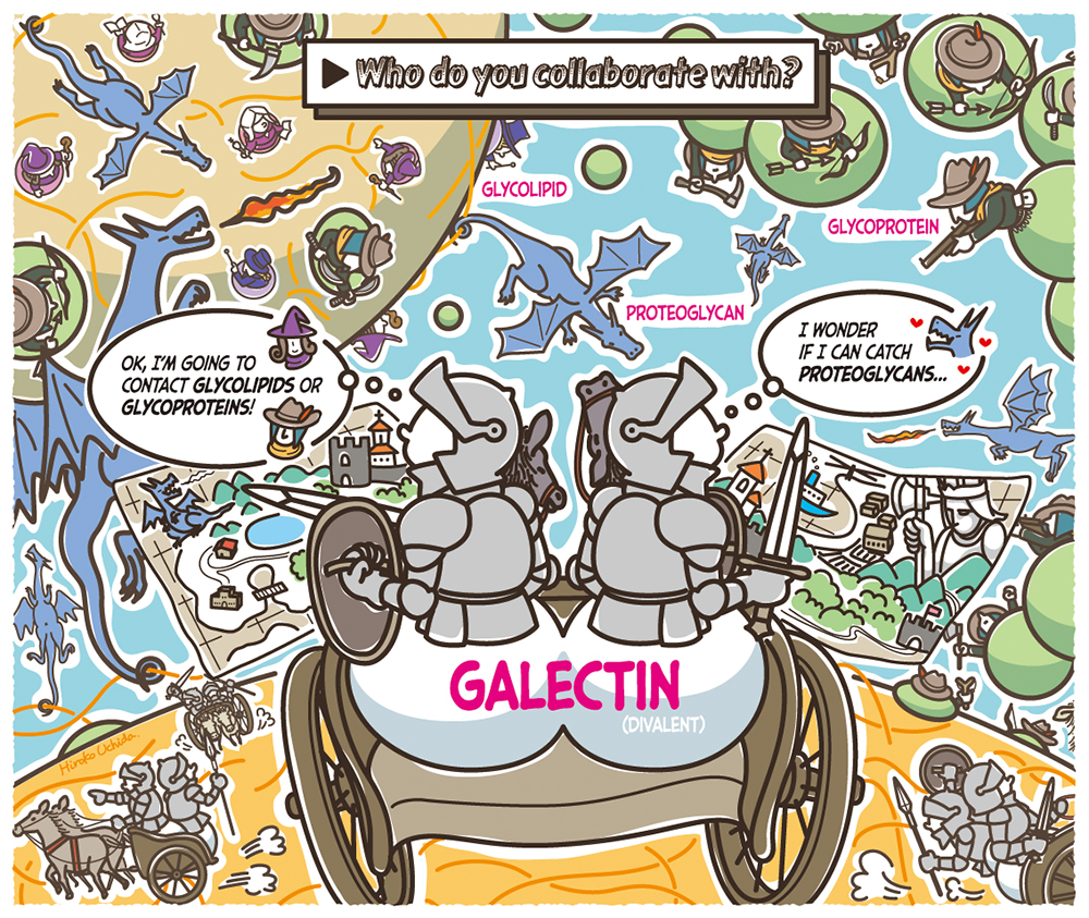
Being a model of lectin interactions, galectin with its weak and loose binding characteristics seems to mediate a wider-than-expected variety of biological activities.
While more than 10 types of galectin have been discovered, all of them bind to lactose and N-acetyl lactosamine as basic recognition units. Therefore, their major targets include lactosyl ceramide and complex-type N-glycan. Various modifications make them recognizable in distinct ways by respective galectins. The individualities of galectin family members (binding forces for respective glycans) have been analyzed in detail using frontal affinity chromatography 2, and these analyses are valuable information sources for understanding galectins 3.
Only a portion of this examination is described here. All galectins exhibit increased binding forces for branched complex-type N-glycans having more than one recognition units. This is attributable to localized high concentrations of the recognition unit. For polylactosamine chains, some galectins (e.g., galectin-3, galectin-9) manifest an increase in binding force with increasing repeats of the recognition unit. These galectins may slide on the chain. Galectin also binds to the low-sulfated region of the keratan sulfate chain.
Thus, galectin-binding glycoconjugates are distributed everywhere in the glycocalyx around the cell, with free glycoproteins occurring everywhere. When dispatched to such sites, galectin serves as “kizuna” (an entity that brings “friendship”) between these so-far-unrelated molecules making use of its cross-linking ability.
Because galectin is insensitive to the aglycon moiety of the glycoconjugate, it does not show preference among glycoproteins, glycolipids, or glycosaminoglycans. It does not matter what work the glycoprotein does. The bindability to galectin and the sustainability of the linkage with galectin depend solely on the structure of the glycan moiety.
What is the level of k-1 for galectin-glycan conjugate? In the past, we attempted to measure the dissociation rate constant of galectin using Biacore.
Glycan containing the recognition unit was flowed through a sensor chip that fixed galectin, but we failed to collect data because of excessively quick binding dissociation.
Sensograms showed that the degree of resonance rose or dropped nearly vertically; therefore, it was impossible to read the half-life of the conjugate (inversely proportional to k-1). Although some studies have reported galectin Kd values for glycan determined using Biacore 4-6, none mentioned rate constants. In some cases, particular glycans were found to dissociate extremely slowly; however, galectin usually releases glycan soon after binding.
On the basis of an unavoidably rough estimation, it was concluded that standalone glycan captured in the binding site seems to leave the site in less than 1 second—a very short time. Hence, it can be described as a fickle protein. In the molecular world, however, even such a short residence may be sufficient to perform a single task.
Studies reported data obtained with galectin passed through a sensor chip that fixed glycan 4-6. In these cases, binding and dissociation take place much more slowly, allowing measurements of k-1 and k+1. Deviating far from the above-described results, what phenomenon occurs depends on whether lectin or glycan is fixed. Because galectin behaves as a divalent entity (the prototype is dimeric and the tandem repeat type has two binding sites, except for galectin-3), multivalent interactions occur under these experimental conditions.
At high fixed-glycan density, rebinding of galectin is likely to occur because diffusion of protein is slower than that of glycan. The low releasability is not a surprise.
These phenomena remind us of cases where galectin has reached the cell surface. In regions where the recognition units are densely distributed, the divalency of galectin serves to extend the retention time and prolong the effect, even if the binding force at each binding site is low.
It has been proposed that a “lattice” involving galectin forms on the cell surface. Although the word “lattice” suggests a firm and stable structure, binding and dissociation occur repeatedly in less than one second everywhere in the lattice. It is reasonable to think that the structures formed with the involvement of galectin are flexible and constantly changing.
In nature, the galectin molecule is a cytoplasmic protein characterized by, for example, the absence of disulfide bonds and glycan. When, for what purpose, and how did it begin operating outside the cell? These questions remain to be answered. Because of the oxidative environment outside the cell, free SH groups will be oxidized or form disulfide bonds, so that force of binding by many galectin molecules will be lost in a relatively short time. The period over which galectin acts seems to be somewhat short.
Its glycan-free structure enables galectin to act freely, unimpeded by extracellular sugar-binding proteins. If entering a glycocalyx, on the other hand, it will be trapped frequently (as it is in an affinity adsorbent). Even so, the galectin will repeat relatively short cycles of escaping the trap and migrating to another glycan. As such, the galectin can initiate various phenomena at anytime in its history; however, it remains unaccountable why the phenomenon is chosen.
Galectin partner selection involves binary uncertainty. First, glycan selection is ambiguous. Second, it does not matter what type of glycoconjugate bears the glycan. This fact could lead to an uncontrollable situation with an ever-increasing number of molecular species influenced by galectin. In reality, however, many studies have reported that galectin is involved in specified events. What program restricts galectin to orderly, rather than chaotic choices? This mystery remains.


