
Kazuo Azuma
Associate Professor, Faculty of Agriculture, Tottori University
Education: Doctor of veterinary medicine
2010 Graduated from Department of Veterinary Medicine, Faculty of Agriculture, Tottori University; licensed as doctor of veterinary medicine. 2013 completed course of the United Graduate School of Veterinary Science Yamaguchi University. September 2013 Assistant professor, Faculty of Agriculture, Tottori University. April 2018 to present, current position. 2017 Incentive Award of the Japanese Society for Chitin and Chitosan. Research interests: Biological function of chitin and chitosan, especially elucidation of function in skin and inflammatory diseases. In veterinary medicine, the relationship between disease and amino acid metabolism and assessment of functional food components, etc., in disease models.
Various biological functions of chitin and chitosan, polysaccharides contained in crab shells, are known. Many studies conducted during the past 50 years have indicated the efficiencies of chitin and chitosan for wound healing. At present, wound dressings made from chitin are also used in the medical field. Here, the wound-healing effects of chitin and chitosan are explained.
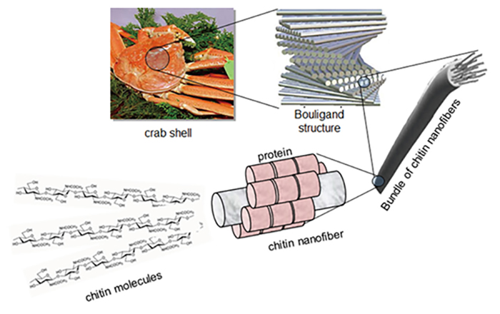
Chitin is a polysaccharide in which N-acetylglucosamine is bound linearly 1. Chitin is contained in crustacean shells, fungal cell walls, and cuticles that cover the surface of invertebrates. In crab shells, fine layers of chitin overlap to form a layer, and the layers overlap each other to form a strong outer shell. Chitosan is obtained by deacetylation of chitin for industrial applications. Chitin and chitosan are polysaccharides that are attracting attention in various fields because of their natural abundance, high biocompatibility, safety, and various biological functions 2.

Chitin and chitosan have various applications mainly in the fields of food supplements. For example, chitosan has an effect to suppress cholesterol adsorption, and glucosamine, which is a monosaccharide of chitosan, is used as a supplement for knee osteoarthritis.
Since around 1970, chitin and chitosan have been known to promote wound healing and are currently commercialized as wound dressings 4. The wound-healing effects are expected to be used not only for the treatment of trauma, but also for the treatment of pressure ulcers in the elderly, the incidence of which has been increasing in recent years. The wound healing promotion effects of chitin and chitosan are summarized in the next section.
The wound healing process can be described in terms of three phases: inflammation, proliferation, and remodeling. Chitin and chitosan have been shown to affect each of these processes 4, 5. Specifically, chitin and chitosan promote the induction of leukocytes and polymorphic leukocytes for foreign body phagocytosis. They also promote the formation of granulation tissue and induce its passage to the proliferative phase. Chitin and chitosan prompt epithelialization, and also induce the release of physiologically active substances such as prostaglandins, which are important for wound healing. Chitin and chitosan enhance platelet aggregation and promote the release of platelet-derived growth factors. Such various growth factors and physiologically active substances induce the proliferations and actions of vascular endothelial cells, fibroblasts, and the like in the wound.
Interestingly, it has been pointed out that chitin and chitosan do not directly stimulate the proliferation of vascular endothelial cells and fibroblasts in vitro. However, the degradation product of chitin and chitosan induces migration activity of vascular endothelial cells. Therefore, chitin and chitosan are considered to contribute to the rapid onset of the inflammatory phase, which is the first stage of wound healing, and its degradation products influence the wound healing process.
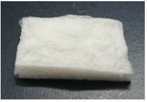
Chitin has been applied as a wound dressing material in clinical medicine because of its wound healing-promoting effect as described above, and also because of its biodegradability and high safety (low antigenicity). In 1989, clinical application for human patients was announced and chitin has been in commercial use to date. For wounds it is used especially for the purposes of “protection”, “maintenance of moist environment”, “promotion of healing”, and “reduction of pain” 6.
The effects of chitin and chitosan are well known not only for humans, but also for animals (veterinary medicine). Minami et al. began investigating applications of chitin and chitosan in veterinary medicine (industrial animals (cattle), companion animals (dogs, cats)) around 1990, and good results have been indicated 4. Based on actual-case experiences, chitin and chitosan have been shown to prevent skin keloidization and are useful for a wide range of wounds. More interesting facts are that chitin and chitosan induced good regeneration of skin, and hair during the healing process. Based on this knowledge, an animal wound dressing using chitin and chitosan was also commercialized in 1992 (so far, it has not been produced , but chitosan cotton are sometimes used for veterinary clinical cases 11.).
In recent years, nanofibers derived from various materials have been produced, including chitin and chitosan. In particular, the group of Prof. Shinsuke Ifuku, Tottori University has succeeded in producing chitin nanofibers from chitin powder using a very simple method of defibration and acid addition 7. The features of chitin nanofibers include a high affinity and dispersibility in water, and stability as a uniform aqueous dispersion.
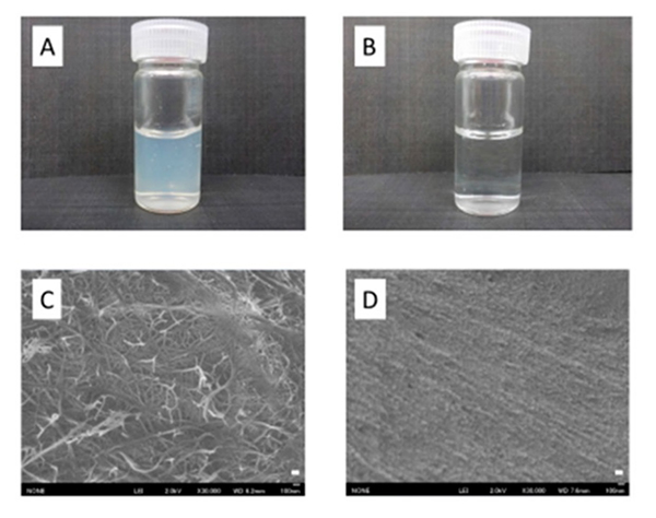
Chitin nanofibers have the physical advantages of nanofibers in addition to the biological functions of conventional chitin, and great expectations are placed on their application. The chitin nanofibers have been experimentally shown to have a wound healing-promoting effect like conventional chitin and chitosan 9. Furthermore, chitin nanofibers have excellent processability, for example, surface-deacetylated chitin nanofibers, in which only the surface of chitin nanofibers is deacetylated, can be produced. Although chitin has not been dissolvable in most solvents in the past, chitin nanofibers form a hydrophilic dispersion, and its applications and processability are dramatically improved: processing of dosage forms such as surface coating and sponge formation is made easy, and it is also easy to produce complexes with other polysaccharides 10.
As described above, the wound healing effects of chitin and chitosan have been studied for about half a century, and have been applied in clinical cases. In addition, chitin and chitosan have various biological functions and are very interesting materials. Research on biological functions and application of chitin nanofibers is being carrying out. Chitin and chitosan are polysaccharides that cannot be missed for future development.


