
Shinji Sakai
Professor, Graduate School of Engineering Science, Osaka University
Appointed as Research Associate and Assistant Professor in the Faculty of Engineering at Kyushu University in October 2002 (Heisei 14). Served as Associate Professor at the Graduate School of Engineering Science, Osaka University from January 2010 (Heisei 22), and was appointed Professor at the same graduate school in April 2016 (Heisei 28). Field of specialization: biochemical engineering.
In the fields of regenerative medicine, drug discovery, and disease modeling, three-dimensional (3D) bioprinting has attracted considerable attention as a technology for arranging cells in three dimensions and reconstructing tissue-specific microenvironments. In this approach, living cell-containing inks (bioinks) are used to fabricate 3D structures. Conventional tissue engineering has generally relied on seeding cells onto prefabricated scaffolds, whereas 3D bioprinting enables the simultaneous placement of cells and materials at desired locations during construct formation, thereby allowing the fabrication of more complex architectures that better resemble native tissues1,2.
In 3D bioprinting, not only printability but also the proliferation and functional expression of encapsulated cells depend strongly on the properties of the bioink. Bioinks are required to provide a highly hydrated environment suitable for cell survival, while also exhibiting sufficient flowability during printing and rapid shape retention after deposition. In addition, the ability to protect cells during printing, support nutrient and oxygen diffusion after printing, and maintain structural stability during long-term culture is also critical. Polysaccharides such as alginate, hyaluronic acid, chitosan, pectin, cellulose-based materials, and sulfated polysaccharides have therefore been extensively investigated as bioink components to meet these requirements. In particular, their biocompatibility, biodegradability, and chemical modifiability make them highly attractive materials for bioink design2,3.
Recent advances in 3D bioprinting have extended the field beyond the simple reproduction of 3D shapes toward the maintenance and induction of cellular functions, the integration of multiple materials, and the rational optimization of printing conditions. Accordingly, diverse gelation mechanisms, multimaterial fabrication strategies, numerical simulations, and artificial intelligence (AI)-assisted approaches are increasingly being incorporated into the design of polysaccharide-based bioinks. Thus, a current understanding of polysaccharide-based 3D bioprinting requires consideration not only of intrinsic material properties but also of crosslinking reactions, printing processes, and digital optimization strategies.
This review focuses on polysaccharide-based 3D bioprinting, particularly extrusion-based bioprinting, and summarizes the characteristics of major polysaccharide materials, crosslinking strategies, and the relationships among printability, gelation behavior, and cellular responses. In addition, representative examples from our own studies, including enzymatic crosslinking systems, visible-light crosslinkable systems, and digital optimization approaches, are introduced to highlight the current status and future challenges of polysaccharide-based bioink design.
Polysaccharides are natural polymers composed of monosaccharide units linked by glycosidic bonds, and they are widely distributed in living organisms and natural resources. Because they generally exhibit high water retention and readily form soft hydrogels, polysaccharides can provide an environment resembling the extracellular matrix (ECM). This characteristic is advantageous for supporting the supply of oxygen and nutrients to cells, as well as the diffusion of metabolic waste productsspan class="sld">4-6. In addition, some polysaccharides interact with cell-surface receptors and growth factors and therefore function not merely as passive supports, but also as microenvironmental materials that influence cell adhesion, migration, proliferation, and differentiation. Accordingly, polysaccharides are important not only as “materials for cell encapsulation” but also as “microenvironmental materials for regulating cellular functions.” Furthermore, because polysaccharides possess a variety of functional groups, including hydroxyl, carboxyl, amino, and sulfate groups, they can be readily modified by introducing photoresponsive or enzyme-responsive moieties and by hybridization with other polymers, thereby enabling control over rheology, crosslinking kinetics, degradability, and biological functionality. For these reasons, polysaccharides represent highly attractive materials for bioink development.
The following sections outline representative polysaccharides that have been used in 3D bioprinting.
Alginate is an anionic polysaccharide derived from brown algae, and its aqueous solution rapidly gels in the presence of divalent cations such as calcium ions. Because this gelation process is mild and straightforward, alginate has long been used as one of the most versatile base materials for bioinks in 3D bioprinting7. However, alginate itself lacks mammalian cell-adhesive motifs, and therefore, when used alone, it does not readily provide gels that sufficiently support cell spreading, proliferation, or tissue organization4. To address this limitation, alginate is often combined with cell-adhesive components such as gelatin or collagen, or modified with adhesive peptides. Thus, while alginate is highly advantageous in terms of printability, it generally requires supplementation to realize its biological functionality.
2-2-2. Hyaluronic AcidHyaluronic acid is one of the principal components of the ECM and is abundantly present in tissues such as skin, cartilage, and vitreous humor. Owing to its high water retention and viscoelasticity, and its involvement in cell migration and differentiation via receptors such as CD44, hyaluronic acid has been widely used in regenerative medicine and tissue engineering8. Whereas alginate has primarily been valued for its printability, hyaluronic acid is particularly important for recreating biologically relevant microenvironments. A major challenge in using hyaluronic acid as a bioink base material is achieving rapid gelation under conditions sufficiently mild for embedded cells. To overcome this issue, hyaluronic acid is frequently used in chemically modified forms, such as methacrylated or phenolated derivatives, that allow photo-crosslinking or enzyme-mediated crosslinking9-12. In this respect, hyaluronic acid is best regarded as a bioink material whose functionality is often unlocked through chemical modification rather than through direct use in its native form.
2-2-3. ChitosanChitosan is a cationic polysaccharide obtained by the deacetylation of chitin and is known to possess antibacterial, hemostatic, and wound-healing-promoting properties13,14. These intrinsic bioactivities make chitosan particularly attractive for applications in which material functionality is directly linked to therapeutic purpose. On the other hand, chitosan is generally more soluble under acidic conditions and is difficult to handle near neutral pH, which presents a major obstacle to its direct use as a bioink base material for cell-laden systems. To address this issue, several strategies have been explored, including the use of neutral-soluble derivatives, Schiff base formation with oxidized polysaccharides, enzymatic crosslinking via phenol introduction, and photo-crosslinking through the introduction of photo-responsive groups15-17. Thus, appropriate chemical design is essential for exploiting the advantages of chitosan in bioink applications.
2-2-4. Plant-derived Polysaccharides: Cellulose, Pectin, Glucomannan, and Related MaterialsPlant-derived polysaccharides have also attracted attention from the standpoint of resource sustainability. For cellulose, one of the most representative plant-derived polysaccharides, studies have examined the gelation of aqueous carboxymethyl cellulose (CMC) solutions by ferric ions18 and the use of methacrylated derivatives as photo-crosslinkable bioink base materials19. Low-methoxyl pectin, which can form ionically crosslinked gels in the presence of divalent cations in a manner similar to alginate, has also been investigated as a bioink base material20. However, CMC and low-methoxyl pectin alone often fail to provide inks with sufficient viscosity for satisfactory shape fidelity. Therefore, small amounts of cellulose nanofibers have been added to enhance shear-thinning behavior and thixotropy, thereby improving printability18,20. In addition, sugar beet pectin has been reported to be useful as a bioink base because it can undergo enzymatic or photo-crosslinking through ferulic acid residues present in its structure21,22. Glucomannan, by imparting dynamic covalent bonding capability and dual-crosslinking properties through chemical modification, has also been explored as a bioink material endowing the bioinks with both good printability and self-healing characteristics15.
2-2-5. Sulfated PolysaccharidesSulfated polysaccharides are frequently found in marine and animal-derived glycans and exhibit the ability to interact with growth factors and cytokines through sulfate groups in their molecular structures. As a result, sulfated polysaccharides such as heparin, chondroitin sulfate, and rhamnan sulfate have attracted attention as functional bioink materials that can contribute to cellular function23,24. Such interactions with bioactive molecules represent a distinctive feature that is less commonly observed in other polysaccharides. In our own work, we have investigated phenol-modified rhamnan sulfate for use in 3D bioprinting and observed characteristic cellular responses reflecting the material's intrinsic bioactivity, suggesting its potential utility in vascularization-related applications25.
As described above, individual polysaccharides possess distinct properties; however, for successful fabrication of 3D constructs, compatibility between the printing mode and the gelation mechanism is essential. In particular, in extrusion-based bioprinting, the ink must remain flowable within the nozzle while maintaining its shape after deposition, even against gravity and the surrounding liquid environment. Accordingly, shear-thinning behavior, yield stress, and rapid gelation are critical design parameters1,2,26.
The gelation mechanisms of bioinks used in 3D bioprinting can be broadly classified into physical crosslinking and covalent crosslinking. Physical crosslinking includes ionic crosslinking of alginate and thermally induced gelation, and these processes are generally simple to operate and have relatively mild effects on cells. However, their mechanical properties may readily change during culture or ion exchange, which can limit long-term stability and the retention of complex structures. In contrast, covalent crosslinking through photochemical or enzymatic reactions tends to produce more stable 3D networks and is therefore advantageous for long-term culture and the preservation of complex shapes. Nevertheless, factors such as photo-initiators, oxidants, and irradiation conditions can influence cellular responses, thereby making reaction control itself part of cytocompatibility design.
In addition to extrusion-based printing, 3D bioprinting also includes inkjet-based, laser-assisted, and vat photopolymerization-based methods such as digital light processing and stereolithography. Because each printing mode requires distinct material properties for successful fabrication, the design of polysaccharide-based bioinks must integrate material characteristics, reactivity, and compatibility with the intended printing process. The next section discusses specific strategies adopted to advance polysaccharide-based bioink systems from this perspective.
As described above, polysaccharides are inherently useful materials, but when used alone they often have insufficient printability or functionality. One of the most representative approaches for overcoming these limitations is chemical modification. Because polysaccharides possess abundant functional groups, the introduction of methacrylate or norbornene groups can impart photo-crosslinkability, while the introduction of phenol groups enables enzyme-mediated crosslinking or photo-oxidative crosslinking7,27-29. In addition, the introduction of aldehyde groups or the exploitation of reactivity with amino groups can be used to design hydrogels with dynamic covalent networks and self-healing properties30. The significance of chemical modification extends beyond the simple introduction of reactivity. Through appropriate modification, it becomes possible to tune mechanical properties, degradability, cellular responsiveness, and even the time window available for printing. Accordingly, chemical modification is a foundational strategy for advancing polysaccharide-based bioinks.
At the same time, increasing the degree of substitution does not always lead to favorable outcomes. Excessive modification may cause reduced water solubility, excessive reactivity, and deterioration of cytocompatibility. Therefore, in bioink design, not only the type of introduced functionality but also the extent of modification must be carefully optimized.
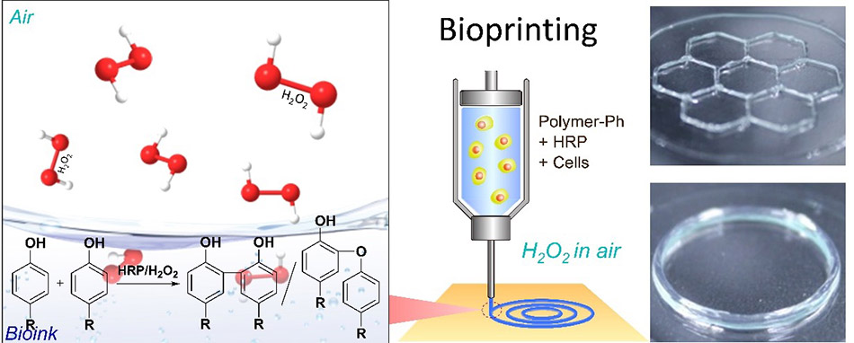
Enzymatic crosslinking has attracted considerable attention as a suitable gelation method for cell-laden bioinks because it can proceed under mild pH and temperature conditions. In particular, the crosslinking of phenol-modified polysaccharides using peroxidases or oxidases enables rapid gelation27 and is highly useful for post-extrusion shape fixation.12 On the other hand, enzymatic systems require careful consideration of substrate and oxidant diffusion, spatial heterogeneity in reaction rates, and the effects of reaction byproducts on cells. Therefore, successful application to bioprinting requires not only the appropriate introduction of enzymes but also the design of the reaction field itself, including the oxidant supply mode and interfacial reaction control.
Photo-crosslinking is advantageous for maintaining fine structural features and enabling selective solidification because of its excellent spatiotemporal controllability. Visible-light-responsive systems are particularly attractive because, compared with ultraviolet light, they are generally less damaging to cells and less likely to induce material degradation. Such systems have increasingly been applied not only to polysaccharide-based bioinks but also to protein-based systems such as gelatin29,31. Whereas enzymatic crosslinking offers strengths in controlling the chemical reaction environment, photo-crosslinking is distinguished by the ability to precisely control irradiation position and exposure duration. However, the choice of initiator, irradiation wavelength, exposure time, and light intensity all influence both crosslinking density and cell viability. Accordingly, the design of photo-crosslinkable bioinks must balance printing speed and crosslinking kinetics, as well as light penetration depth and the presence or absence of auxiliary light-absorbing agents.
It is difficult for a single polysaccharide material to simultaneously meet the requirements of printability, cell adhesiveness, mechanical strength, and high biological activity. For this reason, composite strategies involving proteins, nanofibers, inorganic particles, and other polysaccharides have been widely explored18,20,32,33. The significance of composite formation lies in its ability to compensate for the limitations of individual materials while integrating multiple functions into a single system.
Multimaterial printing, in which multiple inks are selectively used, is also effective for recreating tissue interfaces, controlling spatial cell distribution, and locally tuning mechanical properties34,35. This trend indicates that bioink design has advanced from developing a single “good” ink to the more sophisticated challenge of determining how multiple inks should be spatially organized. In the following section, we introduce representative examples from our own studies.
As a practical example of the enzymatic crosslinking strategy described above, we have continuously investigated systems in which phenol groups are introduced into polysaccharides such as hyaluronic acid, alginate, chitosan, and rhamnan sulfate, followed by crosslinking using horseradish peroxidase (HRP) (Fig. 1)12,25,36,37. These systems proceed under near-neutral conditions that are sufficiently mild for cells and enable rapid gelation, making them highly suitable for extrusion-based bioprinting.
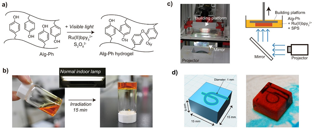
We have also explored visible-light crosslinkable derivatives of polysaccharides. By using visible-light-responsive photoinitiation systems, rapid gelation can be induced under relatively low-invasive conditions, making these systems applicable not only to extrusion-based printing but also to vat photopolymerization-based approaches (Fig. 2)22,31,38. For example, hyaluronic acid-based systems blended with gelatin derivatives have been reported to maintain high cell viability while promoting post-printing structural stability, cell spreading, and the maintenance of stem cell undifferentiation38. Visible-light crosslinking has also been applied to plant-derived polysaccharides such as sugar beet pectin, thereby expanding the range of usable polysaccharide materials22.
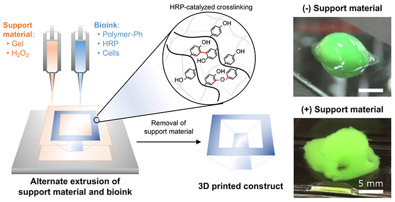
In addition, we have investigated strategies for assisting shape retention in low-viscosity bioinks that are difficult to print on their own by using support materials in combination with the bioink (Fig. 3).39-42 In particular, a method in which a reactive support material and a bioink are alternately deposited enables crosslinking to be induced at the interface immediately after extrusion, thereby contributing to the fabrication of 3D structures with high geometric freedom. This represents an approach in which material design and process design are optimized simultaneously.
Furthermore, we have developed systems capable of switching among multiple inks within a single nozzle43 as well as micromixing technologies that spatially control mixing ratios.44 These technologies provide a foundation for generating interfaces and compositional gradients in polysaccharide-based bioinks. Collectively, these studies illustrate the transition from optimizing single materials to designing multifunctional, multimaterial constructs.
The design of polysaccharide-based bioinks involves many interacting factors, including material composition, viscosity, elasticity, nozzle diameter, extrusion speed, pressure, and cell density. Consequently, purely empirical optimization is inherently limited. This challenge becomes even more pronounced when multiple materials or complex nozzle systems are used, as the parameter space expands rapidly. To address this problem, recent studies have increasingly employed computational fluid dynamics (CFD) and machine learning to predict and optimize printing processes45-47.
CFD is effective for analyzing flow velocity distributions within nozzles, pressure loss, shear stress, and the displacement behavior of dissimilar inks. In our work, we have used numerical analysis in combination with experiments to establish design guidelines for improving switching efficiency in single-nozzle multi-ink systems, with particular attention to the geometry of the junction region43.
Machine learning, on the other hand, is useful for predicting performance under multivariable conditions. We have constructed models that use material composition and printing conditions as inputs to predict post-printing cell viability48. In addition, we have proposed AI-based frameworks for quantifying shape fidelity from printed images, thereby establishing a foundation for objective evaluation of printability and the efficient exploration of optimal conditions49. These approaches should not be viewed as replacements for empirical judgment; rather, they serve as auxiliary tools for efficiently navigating complex parameter spaces.
Nevertheless, these methods still face limitations, including device dependence, material dependence, insufficient training data, and limited extrapolation capability of the models. Accordingly, future progress will require standardized data acquisition protocols, highly reproducible evaluation metrics, and data infrastructures that enable meaningful comparison across different research groups.
Polysaccharide-based 3D bioprinting has evolved into an important technological platform for constructing 3D cell-laden structures by taking advantage of the biocompatibility, biological functionality, and chemical tunability of natural polymers. Alginate, hyaluronic acid, chitosan, plant-derived polysaccharides, and sulfated polysaccharides each possess distinct properties and advantages, and appropriate material selection and molecular design are therefore essential depending on the target application.
In addition, mild crosslinking systems represented by enzymatic and visible-light-mediated reactions, shape-preserving strategies using support materials, and condition optimization aided by simulation and AI are greatly expanding the design freedom and application potential of polysaccharide-based bioinks. As material design, process design, and digital control become increasingly integrated, polysaccharide-based 3D bioprinting is expected to develop into a more reproducible and functional technology that can make substantial contributions to regenerative medicine, drug discovery, and disease modeling.


